September 19, 2024
Histologic Effects Of A New Tool For High-intensity Focused Ultrasound Cyclocoagulation Arvo Journals
Histologic Results Of A Brand-new Tool For High-intensity Focused Ultrasound Cyclocoagulation Arvo Journals Though physiological ramifications of EEG example decline stay to be fully unwinded, these outcomes recommend worsening might serve as a helpful consider discrimination in between control and topics subjected to anxiety waves. To Skinfold understand the influence of ultrasonic application on mind feature, we made use of longitudinal ECoG taping to perform quantitative electrophysiological measurements of neighborhood neural population task from the surface area of the mind. Previous reports have actually described the modifications of quantitative electrophysiology complying with brain injury, consisting of PSD and coherence evaluation at restricted post-injury time factors (summed up in47,48,49,50,51,52). We collected surface area brain activity with implantable electrodes longitudinally from 4 weeks before 6 months complying with ultrasonic treatment used in a very regulated dose and spatial location. In addition, we executed evaluation on the mind signals by quantifying multiple ECoG specifications from 1 to 100 Hz, including PSD, coherence, and decline, which can supply an extensive photo of mind function.Information Analysis
This study will add to the understanding of feasible damages mediated by high-intensity pressure waves. Succeeding studies, employing the devices introduced here, might more thoroughly define the security envelope in ultrasound criterion area. To complement these electrophysiological and behavior strategies, immunohistochemical steps of neuroinflammatory reaction, including microglial and astrocyte reactivity, are obtained in regions throughout the mind. Understanding the relationship in between useful and architectural actions will certainly give an overview in the selection of suitable approaches for safety and security examination of ultrasound while novel standards are been established. Additionally, an understanding of the connection in between ultrasound direct exposure criteria and behavioral and electrophysiological feedbacks will boost our ability to generate brain injury for the objective of diagnosing and treating mTBI. Though the initial results are very motivating, with a level of sensitivity of 100% and specificity of 100% for the ADT technique and 75 and 87.5% for the HSI approach, this research has a number of constraints.High-intensity Focused Ultrasound For The Reduction Of Subcutaneous Adipose Tissue Utilizing Numerous Treatment Techniques
- The majority of occasions had actually settled by 4 weeks and all had actually dealt with at 12 weeks.
- We sonicated 48 mice amount to in this study and did not observe any kind of procedure-related mortality or severe unfavorable events.
- In this paper, we utilized an enhanced SIFT (Scale Invariant Attribute Transform) registration formula for picture enrollment.
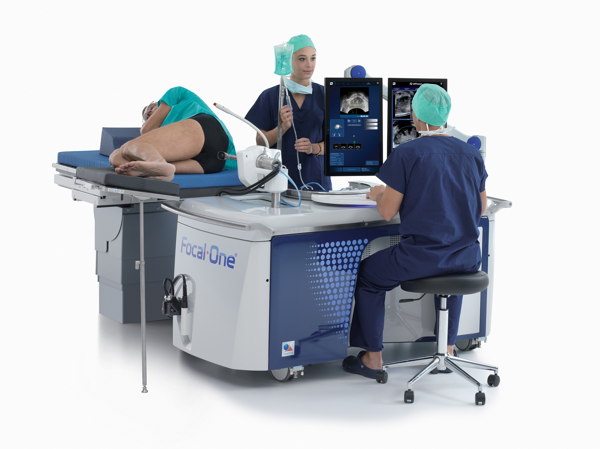
- It consisted of a customer console, the main system, a water closet, an electrical control part, and a healing transducer, as described by [12]
Evidence-based Efficacy Of High-intensity Concentrated Ultrasound (hifu) In Aesthetic Body Contouring
Cells temperature information recorded before, throughout, and after thermal HIFU therapy at the skin surface area, focal zone, and surrounding cells. The yellow-colored mapping shows that the focal zone temperature comes close to 70 ° C, with fast drop-off after treatment. The light blue-colored tracing demonstrates that thermal power is transferred to adjacent cells. Skin temperature (teal, brown, dark blue, and purple tracings) is untouched by the HIFU. A number of different model tools were used during these researches; however, extensive screening and surveillance of power degrees and acoustic outcome parameters showed that the HIFU beam account and outcome power levels were consistent in between models.Tales from the Lab - When Nostradamus got it right about cancer treatment - The Institute of Cancer Research
Tales from the Lab - When Nostradamus got it right about cancer treatment.
Posted: Mon, 08 Feb 2016 08:00:00 GMT [source]
What is the physics behind HIFU?
The device of HIFU healing action takes two types: conversion of power into heat and mechanical cavitation of stress waves in cells. By concentrating the ultrasound waves at a specific area in the body, the energy transformed to heat creates the cells to heat up and kill the cells in the cells.
Social Links